CIRCULATORY SYSTEM OF RABBIT-HEART-STRUCTURE-FUNCTION
-
The circulatory system in animals is the main transport system.
-
In lower animals like protozoa, porifera and cnidaria the transportation of oxygen and nutrients to different organs of the body and expulsion of carbon dioxide and nitrogenous wastes occur by means of diffusion through body surface.
-
However, in higher animals the diffusion of substances does not occur more efficiently due to complexity of the body organisation.
-
A special transport system is essential for transportation of various substances to each and every corner of the body. Consequently the circulatory system is evolved in the animals.
-
In Arthropoda and Mollusca other than Cephalopoda, open type of' circulatory system is present.
-
In open type of circulatory system, definite blood vessels are absent and the flow of blood is confined to sinuses or spaces present in the body.
-
In Cephalopoda (Mollusca), Annelida and Chordata, closed type of circulatory system is developed.
-
In the closed type of circulatory system the blood flows always in definite blood vessels and capillaries. In this system the various nutrients, gases, hormones and wastes are directly supplied to the tissues or mediated by tissue fluids.
-
The circulation is maintained by a pumping centre called heart and blood vessels like arteries and veins.
-
The circulatory system of higher animals is most efficient because of closed circulation.
-
The circulatory system of rabbit is closed type i.e., the blood flows through blood vessels only. The circulatory system constitutes,
i. Heart
ii. Arterial system
iii. Venous system and
iv. Blood
HEART-EXTERNAL STRUCTURE
-
The heart of rabbit is conical, muscular and lies in thoracic cavity between the two lungs.
-
The broader end of heart is towards upper side while the conical end is directed downwards.
-
The heart is slightly towards left side and enclosed by a double walled
pericardium.
-
The inner pericardial layer is called visceral layer while the outer pericardial layer is called parietal layer.
-
A narrow space is left between the two pericardial layers, called pericardial cavity that is filled by pericardial fluid.
-
The pericardial fluid helps in protection of the heart from external shocks and injuries.
- The pericardium is applied to the diaphragm to maintain the position of heart in the thoracic cavity from lungs through pulmonary veins.
- Two aortic arches originate from the ventricles.
- A carotico-systemic aorta originates from the left ventricle while a pulmonary aorta arises from the right ventricle.
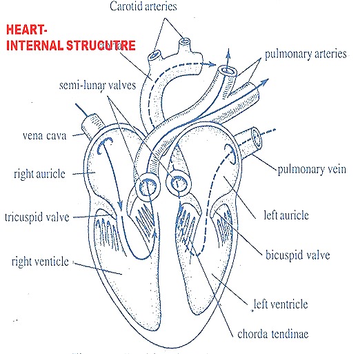
INTERNAL STRUCUTRE OF HEART
-
The two auricles are internally separated by inter-auricular septum.
-
Similarly the two ventricles are internally separated by interventricular septum.
-
The two auricles are communicated with the ventricles by openings called auriculo-ventricular apertures.
-
The two pre-cavals and one post caval open into the right auricle.
-
The opening of posterior vena cava is guarded by a membranous fold

-
called Eustachian valve.
-
The right auriculo-ventricular aperture is guarded by three membranous flaps which constitute tricuspid valve.
-
The tricuspid valve allows the flow of blood from right auricle into right ventricle but prevents the backward flow of blood from ventricle into auricle.
-
The inner surface of the walls of the ventricle is provided with number of ridges called columnae carneae and few conical elevations called the papillary muscles.
-
The free edges of the tricuspid valves are connected to the papillary muscles by white fibres called chordae tendinae.
-
From the left anterior angle of right ventricle a large blood vessel called pulmonary aorta arises that carries blood to lungs.
-
The base of pulmonary aorta is guarded by three semilunar valves which prevent the backward flow of blood from pulmonary aorta into ventricle.
-
The left auriculo-ventricular aperture is guarded by two membranous flaps called bicuspid valve or Mitral valve.
-
The free edges of these flaps are continued into chordae tendinae attached to the papillary muscles of ventricle.
-
At the right angle of left ventricle arises a large aortic trunk called carotico-systemic aorta.
-
The base of carotico - systemic aorta is guarded by three semilunar valves which allow the flow of blood from left ventricle into aorta but prevent the downward flow.
WORKING OF THE HEART
-
The heart of rabbit exhibits rhythmic contractions and relaxations.
-
The contraction is called Systole while the relaxation is called Diastole.
-
The systole and diastole are together referred as Heart beat.
-
The heart beat is initiated by a special node of muscular tissue called sinu - auricular node present on the inner wall of right auricle.
-
The sinuauricular node acts as pace maker and generates certain power in the form of electric impulses.
-
The frequencies of the impulses are governed by nerve fibres.
-
Such impulses are conveyed to the muscles of heart to exhibit rhythmic contractions and relaxations.
-
The impulses are also picked up by another node called auriculo -ventricular node lying on the inter-auricular septum.
-
The impulses are conveyed to Bundle of His and purkinje fibres running
-
through inter - ventricular septum to carry to the wall of ventricles.
-
The right auricle receives impure or de-oxygenated blood from different regions of the body by two superior venae cavae and one inferior vena cava.
-
The left auricle receives pure or oxygenated blood from the lungs by means of pulmonary veins.
-
When the auricles are filled by blood they contract to force the blood into the two ventricles through auriculo- ventricular apertures.
-
When the ventricles are filled with blood, they contract to force the blood into aortic trunks.
-
The backward flow of blood from the ventricles into the auricles is
- prevented by the closure of bicuspid and tricuspid valves. The closure of the bicuspid and tricuspid valves produces first heart beat.
- When the ventricles contract the semilunar valves situated at the base of carotico - systemic trunk and pulmonary aorta are opened.
- As a result the deoxygenated blood from the right ventricle is forced into pulmonary aorta while the pure blood from left ventricle is forced into Aortic arch.
- Now the semilunar valves close to prevent the downward flow of blood from aortic trunks into the ventricles.
- The closure of semilunar valves produces the second heart beat.
- The pulmonary aorta carries deoxygenated blood to the lungs for aeration.
- The aortic trunk supplies pure blood to different organs of the body.
- In different parts of the body the blood gets deoxygenated due to the release of C02.
- The aerated blood from lungs and deoxygenated blood from the tissues enter into the heart to repeat the circulation once again.
