FISH BRAIN ,FROG BRAIN COMPARATIVE ANATOMY
SHARK FISH BRAIN (SCOLIODON BRAIN) FROG BRAIN-COMPARISON
The nervous system is composed of nervous tissue of ectodermal origin. This system is very sensitive and all its parts are structurally connected and functionally integrated. Brain is the anterior part of the central nervous system present in all bilaterally symmetrical vertebrates. Every vertebrate's brain is built in accordance with a single architectural plan. The brain exhibits varying degrees of differentiation and segregation of the sense organs in the head region. The brain of early embryo and of lower vertebrates is almost identical. It is in the form of a swollen anterior end of the neural tube and is known as 'encephalon'. On account of differential growth it gets divided into three 'Primary cerebral vesicles'. These are known as 'FOREBRAIN or PROSENCEPHALON', MIDBRAIN or MESENCEPHALON and HIND BRAIN or RHOMBENCEPHALON.
From the study of brain in different groups of vertebrates, it is evident that the olfactory lobes are more prominent in lower forms are gradually reduced. The brain is enclosed in four ventricles - Lateral ventricles (1&2) or paracoes.the third ventricle or diacoel and fourth ventricle or myelocoel The lateral ventricles communicate with the third ventricle by an aperture Known as 'inter ventricular foramen' or 'foramen of Monro". The third ventricle is connected with the fourth ventricle by a narrow channel called as 'Cerebral aqueduct' or 'Aqueduct of sylvius' or 'Iter'.

The brain is composed with grey and white matter. The grey matter consists of nerve cell bodies with their dendrites and proximal axons. The white matter consists of tracts of medullated fibres connecting the various parts of the brain and carry impulses from and to the spinal cord. A serous fluid the cerebrospinal fluid fills all the ventricles of the brain. It is secreted by the choroid plexus. It is also present in the spaces between the meninges. Here it is absorbed into the blood vessel.

FUNCTIONS OF THE BRAIN

1. Olfactory lobes - Sense of smell
2. Cerebral hemispheres-Seat intelligence and memory.
3. Diencephalon - Controls the general metabolic functions of the body
4. Optic lobes - Sense of vision.
5. Cerebellum - Co-ordinates the movements of voluntary muscles.
6. Medulla oblongata - Controls the involuntary functions of the body.
|
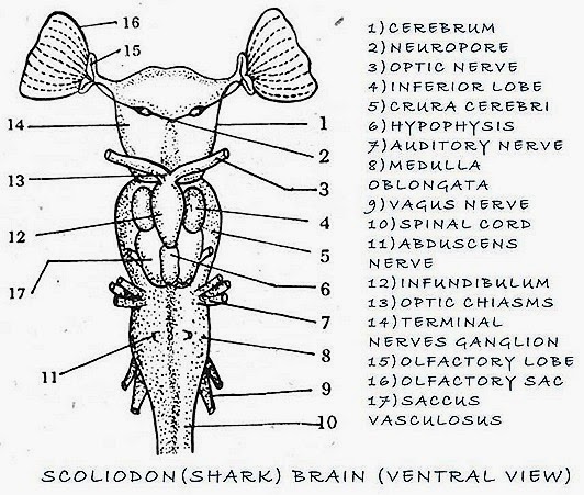
FISH BRAIN (Shark-Scoliodon) |
|
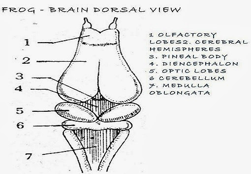
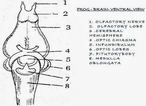
FROG BRAIN |
| 1. The brain lies in a cartilagenous cranium. | 1. The brain lies in a bony cranium. | |
| 2. A single outer membrane - 'Me-ninx primitiva' covers the brain. | 2. An outer thick dura mater, and inner thinner piamater protects the brain so there are two meninges (membranes) | |
| 3. All the ventricles in the brain are spaceous | 3. All the ventricles in the brain are relatively small & narrow. | |
|
SHARK-FISH PROSENCEPHALON |
|
FROG-PROSENCEPHALON |
| 4. The olfactory lobes are highly developed. These help much reliance on smell in detecting the prey . | 4 The olfactory lobes are relatively small. These help much reliance on smell in detecting the prey. | |
| 5. Olfactory lobes extend forward and outward from antero lateral angles of cerebrum. So these do lie side by side. | 5. The olfactory lobes lie side by side and no such differentiation as in shark. | |
| 6. Each olfactory lobe is differentiated into a siende; olfactory tract and a bilobed olfactory bulb. The olfactory bulbs are closely applied to olfactory sacs. | 6. Such a close contact does not exist. | |
| 7. The cerebrum is some what rectangular, large and undivided. | 7. Cerebrum is demarcated into two ovoid cerebral hemispheres by a median fisture. | |
| 8. There is a median neuropore on the ventral sides of the cerebrum for the terminal nerves. | 8. There is no neuropore. | |
| 9. Pallium and corpora striata are poorly developed. | 9. Pallium and corpora striata are well developed. | |
| 10. There is no cerebral cortex. The grey matter is restricted to the lining of lateral ventricles. | 10. Some of the nerve cells of the inner grey matter have migrated to the surface for the formation of cerebral cortex. | |
| 11. Diencephalon is short and narrow and almost fully covered dorsally by cerebrum. | 11. Diencephelon is wide and rhomboi-dal and covered either dorsally or ventrally by cerebrum. | |
| 12. Pineal stalk is long and slender extending forward and upward. | 12. Pineal stalk is very short. It bears distally a knob-like pineal body (tadpole). | |
| 13. Infundibulum consists of a large median lobe associated with a pair of small lateral lobes - 'Lobi inferiores'. | 13. Infundibulum is a median bilobed body. | |
| 14. Median lobe of infundibulum and hypophysis form 'pituitary body'. | 14. Entire infundibulum and hypophysis constitute pituitary body. | |
| 15. A pair of hollow outgrowths 'Sacci vasculosi' present on the sides of hypophysis. These are pressure receptors. | 15. Sacci vasculosi are absent. | |
|
SHARK-MESENCEPHALON |
|
FROG-MESENCEPHALON |
| 16. Optic lobes are highly covered by the cerebellum. | 16. Optic lobes are not covered. | |
| 17. Crura cerebri are largely covered by lobi interiores and sacci vasculosi. | 17. Crura cerebri are slightly covered by pituitary body. | |
|
SHARK-RHOMBENCEPHALON |
|
FROG-RHOMBENCEPHALON |
| 18. Cerebellum is a very large rhom-boidal structure. It divides into three lobes by two 'deep transverse furrows. | 18. Cerebellum is a small transverse band and does not cover any part of the brain. It is undivided. | |
| 19. A pair of irregular, thin -walled sacs 'Corpora restiformia' present on the sides of the anterior part of cerebellum. | 19. There are no corpora restiformia. | |
| 20. Medulla oblongata is partly covered infront by cerebellum. | 20. Medulla oblongata is not covered. | |
| 21. There are three commissures- 'anterior'm lamina terminalis' 'dorsal in front of pineal stalk and 'posterior1 between diencephalon and optic lobes. | 21. These are four commissures -'anterior' in lamina terminalis, 'hippocampal' above anterior commissure 'dorsal infront of pineal stalk and Posterior between diencephalon and optic lobes. | |
| 22. Dorsal commissure is more developed where as posterior commissure is less developed than in frog. | 22. Dorsal commissure is less developed where as posterior commissure is more prominent than in shark. |
