
COELOM AND COELOMODUCTS OF ANNELIDA
The space enclosed in between the two mesodermal layers is called true coelom. The triploblastic animals are classified into three types basing on the presence or absence of this coelom.
1. Acoelomate (wikipedia) : Coelom is absent Ex: Platyhelminthes.
2. Pseudo coelomate (Wikipedia) : The blastocoel is retained in between body wall and alimentary canal as pseudocoelom Ex: Nemathelminthes.
3. Coelomate (Wikipedia) : True coelom is present. These are two types basing on its formation.
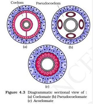
Coelom

a. Schizo coelomate: By splitting of mesoderm layer this true coelom is formed. Ex: Annelida, Arthropoda and Mollusca.
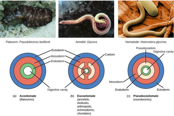
b. Enterocoelcmata: From Archenteron of gastrula two mesodermal pouches will arise. They grow and unite to form true coelom. It is called enterocoelic coelome.Ex : Echinodermata & chordata.
The coelom is filled with coelomic fluid. This coelomic fluid will perform the following functions in Annelid worms.
1. It will keep all the body organs moist.
2. It will allow the peristalasis of the alimentary canal.
3. It works as a fluid skeleton and give turgidity to the body.
4. It will protect the body. Its phagocytic amebocyts will eat away the
pathogenic bacteria which enter into the body.
Coelom ducts : In annelids the body is divided into a number of segments. Each segment will show segmental structures. Coelom ducts and Nephridia are arranged segmentally in these organisms.

1. Coelom ducts : They are derived from mesoderm. They are formed from coelom. They open out on the body through genital pores.They open into the coelom through ciliated funnels called coelomostomes. These coelom ducts work like genital ducts. Because of this these coelomoducts are confined to only few segments in some organisms they secondarily take up excretory function.
2. Nephridia : These are derived from ectoderm. They are excretory structures .They show cilia. They are arranged repeatedly in all segments. They open on the body wall through Nephridio pores.They open into the coelom through funnel like nephrostomes.
Different types of nephridia are present.
a) Protonephridia : These are primitive nephridia. They end blindly in the coelom. They are seen in larval forms of polychates. They bear solenocytes. They resemble flame cells of platyhelminthes.
These protonephridia are present in adult. Polychates like Vanadis, Tomopteris etc.
b) Metanephridia : They open into the body cavity through nephros-tome. It gives a small neck, it will pierce the septal wall and enter the next segment and coil these. There it opens on the body wall through nephridiopore. Thus metanephridia will open on both ends.
Such nephridia are seen in Neries, and members of polycheate, oligo cheate etc.
If these nephridia open on the body wall they are called exo-nephridia. If they are connected with alimentary canal, they are called enteronephric nephridia.
3. Nephromixia : In Hirudinea, oligocheata.and in some polycheates nephridia and coelomoducts are separate.
But in some polycheates nephridia and coelomoducts unite to form nephromixia. Through these excretory matter and reproductive bodies are liberated.
Different types of nephromixia are noticed.
a) Protonephromixia : Ex: Phyllodoce Protonephridia and coelomoducts are united.
b) Meta nephromixia : Ex : Hesione
Metanephridia and coelomoducts are united.
c) Mixo-nephridium : Ex : Arenicola.
Coelomoduct and nephridium will unite as a single duct.
Arenicola
d) Ciliated organs : Ex: Neries.
Coelom ducts are modified as ciliated organs. They are connected with longitudinal muscles and open on the body wall.
Neries
