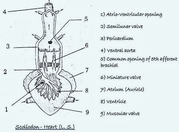
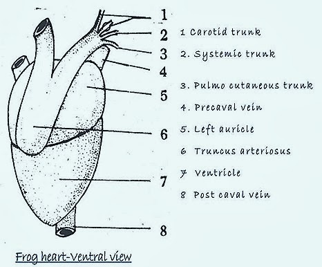
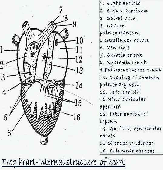
FROG HEART STRUCTURE FISH HEART STRUCTURE –COMPARATIVE ANATOMY
STRUCTURE OF HEART OF SCOLIODON WITH THAT OF RANA-COMPARISION
Scoliodon is a poikilothermc and cartilagenous fish. Rana is also poikilothermic and amphibious animal. In the circulatory system the heart is the most important organ. The blood vascular system in the vertebrates is of closed type. The heart lies in the pericardial cavity of the coelom. It is on the ventral side of the alimentary canal and present anteriorly. In scoliodon the heart is two chambered where as in Rana it is three chambered.

Heart is a pumping organ of blood. From various parts of the body it collect-blood mainly through veins and supplies blood through arteries.

Normally the heart is enclosed by a double walled pericardium which possess pericardial fluid The heart contracts and relaxes rhythmically which facilitate the circulation of blood.

| FISH HEART(SCOLIODON) | FROG HEART(RANA) |
| 1.Heart is approximately pear-shaped. | 1.Heart is approximately pear-shaped. |
| 2.The pericardial cavity is not wide and the pericardium forms double membrane around the heart. | 2.The pericardial cavity is not wide and the pericardium forms double membrane around the heart. |
| 3.The heart is formed of a dorsally placed sinus venosus and ventrally placed two auricle, a ventricle and truncus arteriosus or conus arteriosus. | 3.The heart is formed of a dorsally placed sinus venosus and ventrally placed two auricle, a ventricle and truncus arteriosus or conus arteriosus. |
| 4.The atrium or auricle is two-chambered and lies anterior to the ventricle. Auricles are separated by Inter auricular septum. | 4.The atrium or auricle is two-chambered and lies anterior to the ventricle. Auricles are separated by Inter auricular septum. |
| 5.The auriculo-ventricular valve is membranous. | 5.The auriculo-ventricular valve is membranous. |
| 6.The conu? arteiiosus is incompletely divided by the spiral valve laterally into cavurn aorticum leading to carotid and systemic arches and the cavum pulmocutaneum leading to the pulmocutaneous arch. | 6.The conus arteiiosus is incompletely divided by the spiral valve laterally into cavurn aorticum leading to carotid and systemic arches and the cavum pulmocutaneum leading to the pulmocutaneous arch. |
| 7.The opening of the truncus with valves are arrenged in two transvarse rows | 7.The opening of the truncus with the ventricle is guarded by three semilunar valves arranged in a single row. They devide rruncus into a proximal pylangium and a distal synangium. |
| 8.The walls of the auricle are thick. | 8.The muscular walls of the auricle are thin. |
| 9.The walls of the ventricle are highly muscular. | 9.Same type of ventricle is present. |
| 10.The lips of the bilaminate valves are connected to the inner surface of the ventricle is prominent part of the heart. | 10.The membranous valves are connected to the inner surface of the ventricle by chordae-tendinae. Both auricles and ventricle are essential parts of the heart. |
| 11.The fish heart is venous or branchial heart because it receives deoxygenated blood only. | 11.The frog's heart receives both oxygenated and deoxygenated blood. The deoxygenated blood remain separate in the auricles but get mixed in the ventricle. |
| 12.Blood passes only once through the heart in a complete circuit. | 12.Blood passes through the heart twice in a complete circuit. |
| 13.Such type of arrangement is absent. | 13.The sinus venosus opens into the right auricle through simi-auricular aperture guarded by simi auricular valve which is also known as pace maker. |
| 14.No separate vessel collects oxygenated blood since the heart is venous heart | 14.The oxygenated blood is collected by pulmonary vein from lungs and carries into left auricle. |
