
SYCON SPONGE (SCYPHA) STRUCTURE AND ITS CANAL SYSTEM AND NUTRITION.
ORGANISATION OF SYCON.SPONGE (SCYPHA)
Sycon or Scypha is a typical, small colonial sponge. It belongs to
SYCON-Classification :
Phylum…….Porifera
Class………..Calcarea
Order………Heterocoela
Sycon is widely distributed marine sponge. It is sedentary. It is attached to rocks, shells etc. in shallow sea water.
EXTERNAL CHARACTERS.
1. SIZE AND SHAPE OF SYCON SPONGE: The colony contains groups of cylinders, which are branched. Each cylinder grows three inches in length. All the branched cylinders are connected to a base. At the apex of the cylinder an opening is present called oseulum. Around this opening monaxon spicules are arranged in a circle, called oscular fringe.
2. Colour : The body shows many colours from grey to brown shade.
INTERNAL STRUCTURE OF SYCON:
Sycon is a diplob lastic animal .The body wall is made by two layers
1) Derma 1 layer and 2) Gastral layer. In between them mesenchyme is present.
A) Dermal layer : This layer contains pinacocytes and porocytes.
1. Pinacocytes : These are simple flat, polygonal cells. These are highly contractile. They cover the entire outer body surface of the sponge. Pinacocytes covering the outer body surf from the dermal epithelium and which cover paragastric cavity and form the gastral epithelium.
2. Potocytes :These are tubular cells distributed among the pinacocytes. They form the openings on the dermal layer.
B) Gastral layer : it shows choanocytes and epithelial cells.
Qvanocytes : These are round cells. They show big nucleus A long flagellum s rises from each cell. At base of the flagellum a protoplasmic collar is present. The action of flagellum brings in water. This cell is useful in digestion, respiration and other functions.
C) Mesenchyme : It is present between dermal and gastral layers. It contains amoebocytes. They are many types.
1) Scleroblasts : The amoebocytes secrete skeleton. Scleroblasts are of three types :
1) Calcoblasts . Scleroblasts that secrete calcareous spicules.
ii) Silicoblasts . Scleroblasts that secrete silicious spicules
iii') Spongioblasts . Scleroblasts that secrete spongin fibres.
2) Chromocytes: Amoebocytes with pigment and give colour to the body.
3) Thesocytes : These cells contain reserve food material.
4) Archeocytes : These are big in size. They give rise to sex cells.
5) Myocytes : These are highly contractile cells. They are arranged circularly around the osculum arid other openings. They guard and regulate the apertures.
6) Gland cells : They are attached to the surface of the sponge. They produce slime.
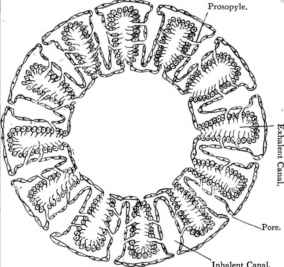
CANAL SYSTEM IN SYCON SPONGE:
Sycon shows syconoid type of canal system.
a) Dermal ostla : On the body of the sycon dermal ostia are present. They open into incurrent canals
b) In current canals : These are narrow canals. These are covered inside by epethelial cells. They show some openings here and there called prosopyles.
c) Radial canal: The body wall is folded. In these foldings radial canals are present at regular intervals. Prosopyles open into these Radial canals.
In between the two successive radial canals a tubular space, called incurrent canal, is present. Thus radial canals and incurrent canals are arranged alternately.
The radial canals are lined with flagellated cells The flagellar action brings water into the body.
d) Excurrent chambers : The radial canal opens into excurrent chamber through apopyle openings. This chamber is lined by epithelial cells. It opens into spongocoel.
e) Spongocoel : In the centre of the body of sycon a narrow cavity is present called spongocoel. It opens out through osculum.
Transverse Section (T.S.) of Sycon (TS of Sycon)
NUTRITION IN SYCON SPONGE:
a) Food : The food of sycon is small minute bacteria, diatoms, protozoans etc. The food particles come into the sponge along with water current.
b) Digestion : The digestion is intracellular. The food particles are usually captured by the choanocytes. Digestion takes place in the choanocytes. The digested food is passed to other cells. The reserve food is stored in the form of fats, glycogen and Droteins in the thesocyte.
The undigested marter sent out through osculum along with excurrent water current.
SYCON SPONGE-SPICULES :
In sycon the skeleton contains calcareous spicules. These are of the following types,
(i) Large one rayed needle like Monaxon spicules,
(ii) Simple monaxon spicules project from dermal layers on the walls of the radial canals,
(iii) Triaxon spicules are present on the walls of spongocoel.
(iv) Tetraxon spicules are present on the walls of spongocoel.
RESPIRATION : Respiratory organs are absent in sycon. Respiration is by simple diffusion, between the cells of sponge and water.
EXCRETION : In sycon excretory organs are absent. It is done by diffusion. Some people say the excretory wastes will go out of the body through excurrent water.
REPRODUCTION IN SYCON SPONGE: it is carried on by asexual and sexual methods. Budding is the common asexual method.
Sexual reproduction is carried on by the development of sperms and ova. Fertilisation is internal. In the life history Amphiblastula larva is seen .