FROG-REPRODUCTIVE SYSTEM-FISH- REPRODUCTIVE SYSTEM-COMPARISION
MALE URINOGENITAL SYSTEM OF SHARK FISH (SCOLIODON) AND FROG(RANA)AND FEMALE REPRODUCTIVE SYSTEM IN SHARK AND FROG-COMPARATIVE ANATOMY
Shark is a marine cartilagenous fish, it is also known as scoliodon which is a poikilothermic animal. Frog is an amphibious animal. Both the animals have been included in Anamniota Frog is also poikilothermic animal.
Reproductive system consists of gonads and genital ducts. The gonads are the essential reproductive organs. In male the gonads are the 'testes' or 'spermaries' and 'ovaries' in the female. The gonads are mesoder¬mal in origin and develop from a pair of genital ridges of the embryo. The genital ridges are the folds of coelomic epithelium lying on the medial sides of the embryonic kidneys. When fully formed, the gonads hang from the dorsal body wall of the double folds of peritoneum called as 'Mesorchia' in the case of testes and 'mesovaria' in the case of ovaries. The gonads produce sex cells or gametes. They are known as 'spermatozoa or sperms' in the male and 'ova' in the female. The gonads also secrete some important hormones.
The genital or reproductive ducts are developed for transporting the gametes produced by the gonads. These are known as ‘Vasa deferentia'
in the male and 'oviducts' in the female.
There is an intimate relation present between the ureters and genital ducts particularly in the male. Hence the excretory and reproductive systems are collectively reffered as urinogenital system.


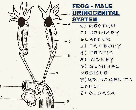
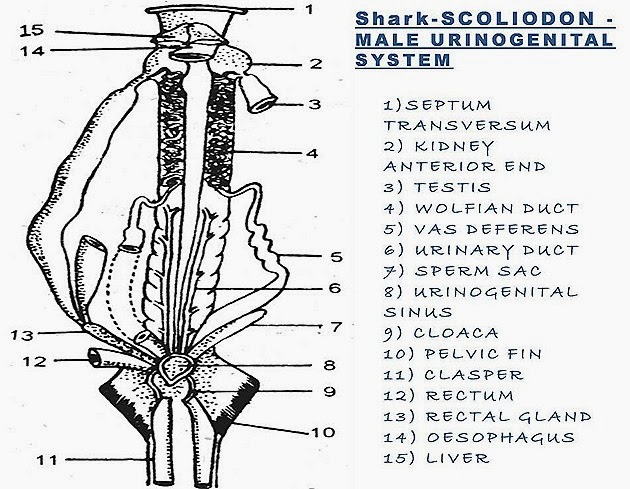
MALE REPRODUCTIVE SYSTEM OF SHARK FISH(SCOLIODON)- MALE REPRODUCTIVE SYSTEM FROG(RANA)-SIMLARITIES AND DIFFERNCES
| SHARK-MALE - URENOGENITAL SYSTEM | FROG -MALE - URENOGENITAL SYSTEM |
| L. Testes are very long and ribbon like. | 1. Testes are small and rod-like. |
| 2. Testes are attached to the kidneys anteriorly. | 2. Testes are attached to the kidneys above the middle region with mesorchium. |
| 3. Testes are connected with the rectal gland by epigonal organs. | 3. The epigonal organs are absent. |
| 4. Vasa efferentia leave the testis at its anterior end. | 4. Vasa efferentia leave the testis alone its inner border. |
| 5. These open into the wolffian duct. | 5. These enter the kidney and open into .the Bidder's canal which drains into the wolffian duct. |
| 6. Wolffian duct is differentiated into an anterior narrow closely convoluted epididymis and the posterior wide less convoluted vesiculus seminalis. | 6. Wolffian duct is notdefferentiatedint< the parts except having a small seminal vesicle near its beginning. |
| 7. Wolffian ducts act only as genital ducts. | 7. Wolffian ducts act as both urinary and genital duct. Hence they are known as urinogenital ducts. |
| 8. A pair of club-shaped sperm sac open into the urinogenital sinus | 8. Sperm sacs are absent |
| 9. The copulatory apparatus comprising siphons and claspers. | 9. Copulatory apparatus is absent. |
| 10. Sperms are released into the genita iuct of female, hence fertilization is internal. | 10. Sperms are released over the eggs tithe fresh water, hence fertilization is external. Sperms are released as milt' |
| 11. There are no fat bodies attachec to the testes. | 11. There is a large branched fai-bodv attached to the anterior end of ead testis. |
| 12. There is a single urino-genital papilla. | 12. Urino-genital papillae are paired. |
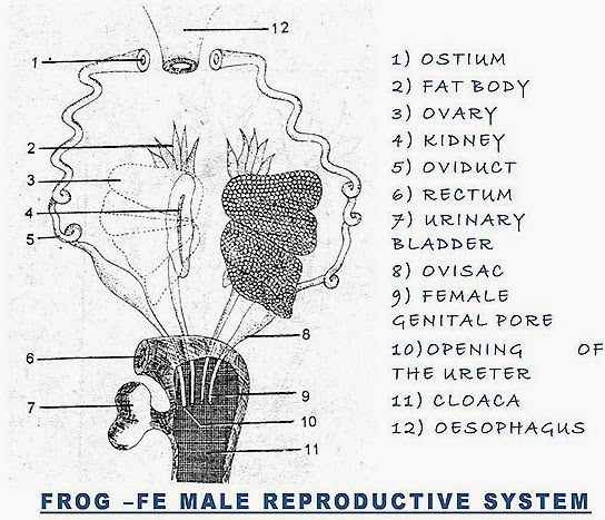
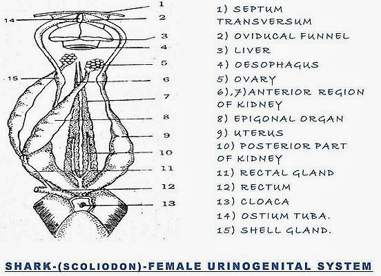
FEMALE REPRODUCTIVE SYSTEM OF FISH(SCOLIODON-SHARK)- VS -FEMALE REPRODUCTIVE SYSTEM FROG(RANA)


| SHARK -FEMALE - REPRODUCTIVE SYSTEM | FROG -FEMALE - REPRODUCTIVE SYSTEM |
| 1. Ovaries are small, tabulated bodies located just behind the base of the liver. | 1. Ovaries are large, hollow, lobed sacs laying on the ventral surface of the kidneys. |
| 2. Ovaries are connected with rectal glands by long epigonal organs. | 2. Epigonal organs are absent. |
| 3. Oviducts (Mullerian ducts) are long but not convoluted. | 3. Oviducts (Mullerian ducts) are very long and greatly convoluted. |
| 4. Oviducts converge and unite in front of the ovaries leaving a slit 'ostium tubae! The oviducal funnels are on either side of ostium tubae. | 4. Oviducts converge infront of the ovaries but do not unite. Each oviduct has its own ostium at the lip of oviducal funnel. |
| 5. The oviducts possess a small enlargement behind ovaries This enlargement is called shell gland. | 5. There is no shell gland |
| 6. The oviducts are expanded to form large uteri in the region of renal part of kidneys | 6. The oviducts are expanded to form small ovisacs behind the kidneys. |
| 7. The oviducts join to form a median sac-'vagina' which opens into the cloaca. | 7. The oviducts independently open into the cloaca. The vagina is absent. |
| 8. There are no fat bodies. | 8, There is a large fat body attached to the anterior end of each ovary. |
| 9 Since the fertilization in internal, there is no question of releasing the egg out side the body | 9. The mass of eggs is called "spawn* which is released outside the body Fertilization is external. |
