
PALAEMON-(PRAWN)- NERVOUS SYSTEM-SENSE ORGANS
The nervous system of Palaemon resembles that of annelidas, it shows
1. Central nervous system.
2. Peripheral nervous system.
3. Autonomic nervous system.
- Central nervous system(CNS): The central nervous system of Palaemon is made up of three main components. It includes a pair of cerebral ganglia, a pair of circum esophageal connectives and a double ventral nerve cord :

i) Cerebral ganglia(Brain):
- Located anteriorly, just above the junction of the oesophagus with the cardiac stomach.
- Appears as a white, bilobed mass.
- Often referred to as the "brain" of the prawn.
- Acts as the primary control center for sensory processing and motor coordination.
ii) Circum oesophageal connectives :
- These are paired nerve cords that originate from the postero-lateral regions of the cerebral ganglia.
- They descend around the oesophagus and connect to the ventral nerve cord.
- Each connective contains a small commissural ganglion that gives rise to the mandibular nerve , supplying the mandible.
iii) Double Ventral nerve cord :
- It lies in the mid-ventral line of the body, it bears seventeen pairs ganglia.
- Tare anterior eleven pairs of ganglia belong to the cephalothorax and are fused to form a single and large mass called the Cephalo thoracic ganglionic mass.
- The posterior six pairs of ganglia of the nerve cord belong to the abdomen and they remain separate.
- The sixth and abdominal ganglion is larger than others.
2. Peripheral nervous system : It contains paired nerves that arise from the central nervous system to innervate various parts of the body.
a)Nerves Arising from the Brain (5 pairs) :
- Optic Nerves – extend through the eye stalks and supply the compound eyes.
- Ophthalmic Nerves – arise behind the optic nerves and supply the ocular muscles within the eye stalks.
- Antennulary Nerves – originate antero-ventrally from the brain and innervate the antennules; also give off the statocystic nerve to the statocyst.
- Antennary Nerves – arise from the ventral side of the brain and supply the antennae.
- Tegumental Nerves – arise posterior to the antennary nerves and innervate the labrum.
b) Nerves from the Cephalothoracic Ganglionic Mass (11 pairs) :
Eleven pairs of nerves originate from the cephalo thoracic ganglionic mass.
- Mandibular Nerves (1 pair) – supply the mandibles.
- Maxillulary Nerves (1 pair) – innervate the maxillulae.
- Maxillary Nerves (1 pair) – supply the maxillae.
- Maxillipede Nerves (3 pairs) – supply the maxillipeds.
- Walking Leg Nerves (5 pairs) – each pair supplies one pair of walking legs.
c) Nerves from Abdominal ganglia:
- The fifth abdominal ganglion gives only two pairs of nerves.
- The sixth abdominal ganglion gives six pairs of nerves.
3. Autonomic nervous system:
The autonomic nervous system regulates involuntary functions such as digestion and visceral muscle movement.Composed of two visceral ganglia :
- Anterior Visceral Ganglion : connected via short transverse connectives to the commissural ganglia of the circumoesophageal connectives.
- Posterior Visceral Ganglion : provides two pairs of nerves to the muscles lining the walls of the oesophagus and stomach.
SENSE ORGANS OF PALAEMON
Palaemon shows different sense organs. They are
- STATOCYSTS,
- TACTILE SETAE,
- OLFACTORY SETAE,
- COMPOUND EYES.
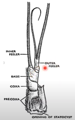
1) STATOCYSTS :
- A pair of statocysts is present in the precoxa of each antennule.
- Structure:
- Round sac, approximately 1–1.5 mm in diameter .
- Outer opening covered by a thin integument.
- Inner cavity lined with an oval ring of receptor setae and filled with sand grains (statoliths) .
- Receptor setae have:
- A swollen base attached to the sac wall via a membrane.
- A slender shaft with fine bristles above a bent region.
- Supplied by the statocystic nerve .
Working Mechanism :
- Statoliths (sand particles) press against specific receptor setae when the animal’s position changes.
- This pressure stimulates nerve fibers, sending signals to the brain to adjust posture and maintain equilibrium.
A) Statocyst in antennule B) Single receptor setae
C) Tactile setae D) Olfactory setae E) T.S. of Statocyst
1) Precoxa
2) Statocyst
3) Antennary nerve
4) Sensory hair
5) Statoliths
6) Cuticle
7) Blade
8) Root
2) TACTILE SETAE - Touch Receptors:The tactile setae are seen on the antennae and on other parts of the body.
A tactile setae has two parts.
- Stout Proximal segment : Articulating with the integument by a flexible arthrodial membrane.
- Distal segment (blade): slender and sensitive to touch; bears two rows of barbs.
3) OLFACTORY SETAE - Smell Receptors :
- Located on the middle small feeler of each antennule.
- Each seta consists of:
- A stalk embedded in the cuticle.
- A blade that receives olfactory stimuli.
- Innervated by a small nerve branch from the antennary nerve .
4) Compound EYES - Visual Organs:
- A pair of compound eyes , mounted on movable stalks , located near the base of the rostrum.
- Each eye-stalk has two segments:
-
-
- Proximal segment – attached to the body integument.
- Distal segment – carries the compound eye at its tip.
-
Structure of the Eye:
Each compound eye comprises numerous ommatidia , arranged radially.

1) Dioptric part:
- Covered by a transparent cornea .
- Corneal lens is biconvex, secreted by a pair of comeageal cells .
- Beneath these lie crystalline cone cells (four vitrellae cells) .
- Collectively, these form the dioptric part of the ommatidium.

2) Retinal part:
- Below the dioptric part a long, refractile rod, the rhabdome is present.
- It is secreted and surrounded by seven long cells, the retinulae.
- The end of each retinal cells is prolonged into a nerve fiber that joins the optic nerve.
- The rhabdome and the retinulae from the retinal part of the ommatidium.
- Each ommatidium is covered by pigmented cells which will separate it from the nearby ommatidium.
Function:
- During bright light, ommatidia are completely covered by the pigment cells.
- The ommatidia are separate from one another.
- Thus many images are developed. Such-an image is called the opposition or mosaic image and the eye is said to have a mosaic vision.
- During dim light pigment cells separate apart and expose the ommatidia. With the result, the rays of light entering several adjacent corneae will overlap The image is formed by overlapping points of light.
- Such an image is known superposition image.