
CHOANOCYTES –COLLAR CELLS
CHOANOCYTES IN PORIFERA OR FLAGELLATED CELLS IN SPONGES
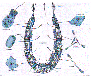
There are different types of cells presents in sponges.The gastral layer of sponge contains a special type of cells called as choanocyte cells. These are also called flagellated cells.
Each cell is big. It is oval in shape. It has a big nucleus. It shows contractile vacuole and food vacuole. From a basal granule a flagellum arises. At the base of the flagellum a protoplasmic collar is present. Hence it is also collar cell.
In Sycon, these cells are restricted to radial canals. They are called flagellated chambers. By the action of flagelia water current is brought in. The incurrent water brings in food particles and 02. These food particles are filtered by the collar of the choanocyte. These food particles may be digested by the choanocyte or may be given to the amoebocyte.
Additional Note on Cellular Structure in Sycon Sponges
There are several types of cells present in sponges, each performing a unique physiological function that ensures survival and filtration efficiency.
The gastral layer (inner layer) of the sponge contains a distinctive type of cell known as the choanocyte, or collar cell, which is characteristic of all members of the phylum Porifera.
Each choanocyte is relatively large and oval in shape, containing a prominent nucleus, a contractile vacuole, and a food vacuole.
From a basal granule, a single flagellum extends outward, surrounded at its base by a thin protoplasmic collar hence the term collar cell.
This structure allows the cell to generate water currents while simultaneously trapping suspended food particles.
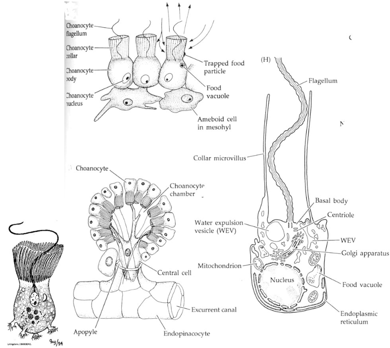
In Sycon, choanocytes are not scattered throughout the sponge body but are restricted to the radial canals, forming what are known as flagellated chambers.
By the rhythmic beating of their flagella, these cells draw water inward through the ostia and canals. The incoming current carries oxygen and microscopic food particles, which are trapped by the collar and filtered effectively.
Captured particles are either digested within the choanocyte itself or transferred to neighboring amoebocytes for intracellular digestion and nutrient distribution.
This cellular arrangement highlights the remarkable coordination between choanocytes and amoebocytes, demonstrating how even primitive multicellular organisms maintain specialized systems for feeding, respiration, and internal transport.